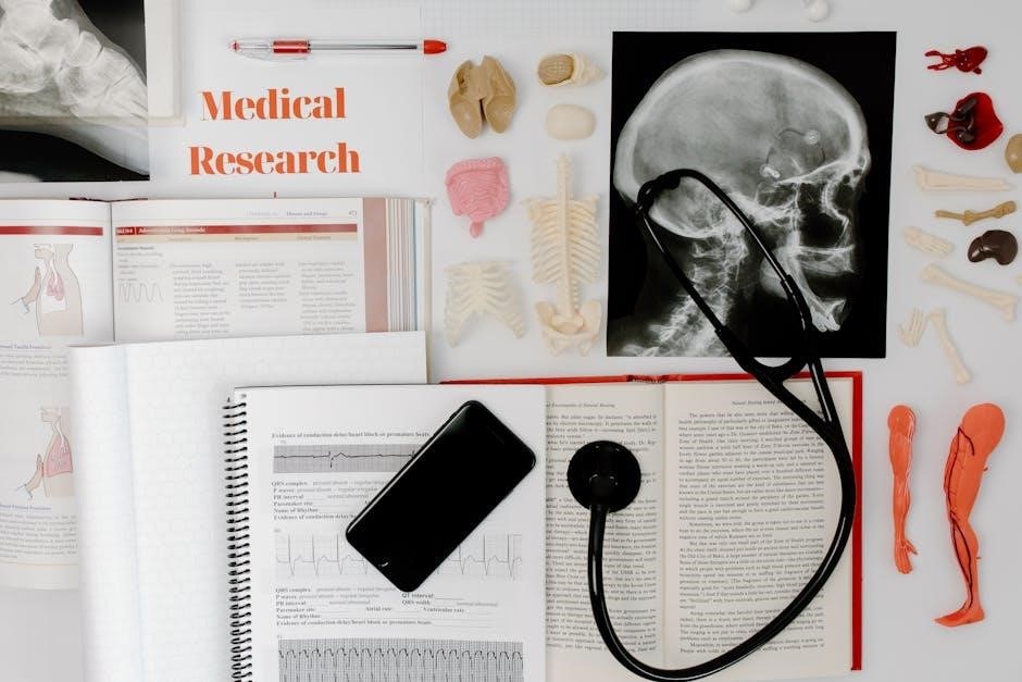
Welcome to the Anatomy & Physiology Laboratory Manual, a comprehensive guide designed to complement your textbook and enhance hands-on learning. This manual aligns with two-semester courses, providing detailed exercises, visual aids, and practical activities to explore anatomical structures and physiological processes. With a focus on observational and laboratory skills, it supports students in healthcare programs, offering a structured approach to understanding the human body’s complexities. Perfect for classroom and lab use, it integrates seamlessly with online resources like Mastering A&P for a holistic learning experience.
1.1 Purpose and Scope
The Anatomy & Physiology Laboratory Manual is designed to provide students with a hands-on, interactive learning experience, complementing classroom instruction and textbook content. Its primary purpose is to guide students through structured lab exercises that reinforce anatomical and physiological concepts. The manual covers a wide range of topics, from basic anatomical terminology to complex organ system studies, ensuring a comprehensive understanding of the human body. It is tailored for two-semester courses and aligns with various teaching materials, including online platforms like Mastering A&P. The scope includes practical activities, dissections, and experiments to enhance observational and critical thinking skills, preparing students for careers in healthcare.
1.2 Key Features of the Manual
The Anatomy & Physiology Laboratory Manual offers a variety of interactive and hands-on activities designed to enhance learning. It includes high-quality illustrations, step-by-step dissection guides, and activities tailored to different learning styles. The manual features pre-lab videos, clinical application questions, and critical thinking exercises to reinforce concepts. With versions for main, cat, and fetal pig dissections, it caters to diverse lab needs. Additionally, it integrates with Mastering A&P, providing online resources like practice quizzes and flashcards. This comprehensive guide supports students in developing practical skills and a deeper understanding of human anatomy and physiology.
Laboratory Safety and Procedures
This section outlines essential safety protocols and procedures for conducting laboratory exercises. It emphasizes proper use of equipment, personal protective gear, and emergency response to ensure a secure learning environment.
2.1 General Safety Guidelines
Adhering to safety protocols is crucial in the anatomy and physiology lab. Always wear personal protective equipment (PPE) such as gloves and goggles when handling specimens or chemicals. Familiarize yourself with emergency exits, fire extinguishers, and first aid kits. Avoid ingesting or touching potentially hazardous materials. Follow proper hygiene practices, including washing hands before and after lab activities. Be mindful of sharp instruments and electrical equipment. Dispose of biological waste and chemicals according to lab regulations. Attend all safety briefings and adhere to instructions from lab instructors. Preparedness and responsible behavior ensure a safe and effective learning environment.
2.2 Proper Use of Laboratory Equipment
Properly using laboratory equipment is essential for safety and accuracy. Begin by inspecting equipment for damage or wear. For microscopes, clean lenses with soft cloth and use immersion oil as needed. Handle dissection tools carefully, ensuring blades are sharp and securely stored. When using balances or measuring tools, calibrate them before use. Always follow manufacturer instructions and lab protocols. After use, clean and return equipment to its designated storage area. Never share personal equipment or use shared tools without proper sterilization. Familiarize yourself with equipment functions through provided guides or instructor demonstrations to ensure efficient and safe operation.

Overview of the Human Body
This section introduces foundational anatomy and physiology concepts, exploring the structure and function of the human body. It provides a comprehensive overview of body systems and their interconnections, focusing on how they maintain homeostasis and support overall health. Through detailed descriptions and visual aids, students gain a holistic understanding of the body’s organization and physiological processes.
3.1 Anatomical Terminology
Anatomical terminology forms the foundation of understanding the human body. This section introduces standardized terms used to describe body structures, directions, and positions. Students learn to identify and use correct terminology for regions like the abdominal, thoracic, and pelvic cavities. Key concepts include proximal-distal, anterior-posterior, and superior-inferior orientations. The manual provides exercises to master these terms through labeling diagrams and interactive activities. Clear definitions and visual aids ensure a strong grasp of terminology, essential for accurate communication in healthcare and scientific settings. This knowledge is applied in subsequent sections to identify organs and systems effectively.
3.2 Body Systems Overview
This section provides a concise overview of the human body’s major systems, including the skeletal, muscular, nervous, digestive, circulatory, respiratory, urinary, and reproductive systems. Each system’s primary functions and interconnections are explored to illustrate how they maintain homeostasis and overall health. The manual includes detailed diagrams, flowcharts, and exercises to help students visualize and understand the integrated roles of these systems. Activities focus on identifying organs, their locations, and interactions, reinforcing a holistic understanding of human anatomy and physiology. This foundational knowledge prepares students for more detailed studies in subsequent chapters.
Cellular and Tissue Studies
This section introduces fundamental concepts of cells and tissues, essential for understanding human anatomy. It includes microscopy techniques, tissue identification, and hands-on activities to explore cellular structures and their roles in forming organs.
4.1 Microscopy Techniques
Mastering microscopy is essential for exploring cellular and tissue structures. This section covers light microscopy, electron microscopy, and fluorescence microscopy, detailing their applications and limitations. Students learn to prepare slides, including fixation, staining, and sectioning, to visualize cellular details. Hands-on exercises emphasize proper microscope handling, focusing, and specimen observation. These techniques are crucial for identifying cell types, understanding tissue organization, and analyzing physiological processes. Practical activities align with textbook content, reinforcing concepts through direct observation and experimentation, making microscopy a cornerstone of anatomy and physiology studies.
4.2 Tissue Identification
Tissue identification is a critical skill in anatomy and physiology, focusing on recognizing and differentiating the four primary tissue types: epithelial, connective, muscle, and nervous. Through microscopy, students analyze tissue samples to observe cellular arrangements, shapes, and specialized features. Practical exercises involve identifying tissues using histological slides, including staining techniques like H&E, to enhance visibility. This hands-on approach helps students correlate microscopic structures with their physiological functions, reinforcing understanding of tissue organization and their roles in maintaining body systems. These skills are essential for advanced studies in histology and pathology.
Skeletal System
The skeletal system explores bone structure, classification, and function. It focuses on the axial and appendicular skeleton, emphasizing the role of bones in movement and body support.
5.1 Bone Structure and Classification
This section examines the detailed structure of bones, including their composition and functions. Bones are categorized into long, short, flat, irregular, and sesamoid types, each serving unique roles. Long bones, like the femur, support body weight and enable movement, while flat bones, such as the skull, provide protection; Short bones, found in the wrist, allow limited motion, and irregular bones, like vertebrae, have complex shapes for specialized functions. Understanding bone classification and structure is essential for studying the skeletal system’s role in movement, support, and protection. This section also covers bone markings and surface features, enhancing your ability to identify and analyze skeletal components in lab settings.
5.2 Axial and Appendicular Skeleton
The human skeleton is divided into two main divisions: the axial skeleton and the appendicular skeleton. The axial skeleton includes the skull, vertebral column, ribs, and sternum, forming the body’s central framework and protecting vital organs like the brain and spinal cord. The appendicular skeleton comprises the upper and lower limbs, shoulders, hips, and pelvis, enabling movement and locomotion. This section explores the structural and functional differences between these two systems, highlighting how they work together to provide support and facilitate mobility in the human body. Understanding their roles is crucial for analyzing skeletal anatomy in laboratory settings.
Muscular System
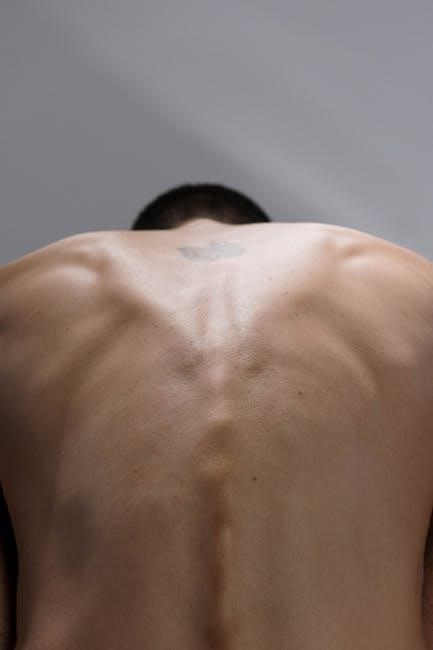
The muscular system consists of over 600 muscles, enabling movement, stability, and support for the body. It includes skeletal, smooth, and cardiac muscles, each with unique functions and structures. This section provides a detailed exploration of muscle anatomy, physiology, and their roles in maintaining homeostasis and facilitating voluntary and involuntary movements.
6.1 Skeletal Muscle Anatomy
Skeletal muscles are voluntary muscles attached to bones via tendons, enabling movement and locomotion. They consist of long, multinucleated muscle fibers packed with myofibrils. Each myofibril contains sarcomeres, the functional units responsible for contraction. Skeletal muscles are organized into fascicles, surrounded by connective tissue, and innervated by motor neurons. Laboratory exercises involve identifying muscle origins, insertions, and actions, as well as exploring histological preparations to visualize muscle fiber structure. Understanding skeletal muscle anatomy is crucial for grasping movement mechanics and the musculoskeletal system’s role in body function.
6.2 Major Muscle Groups
The human body is divided into several major muscle groups, each responsible for specific movements and functions. These include the anterior and posterior muscle groups of the upper and lower limbs, as well as the abdominal and back muscles. The anterior muscles, such as the biceps and quadriceps, facilitate flexion, while the posterior muscles, like the triceps and hamstrings, enable extension. Laboratory exercises involve identifying and palpating these groups, exploring their origins, insertions, and actions. Understanding these muscle groups is essential for comprehending movement, posture, and overall musculoskeletal function.
Nervous System
The nervous system includes the brain, spinal cord, and nerves, controlling bodily functions like movement and sensation. This section explores neural structures, nerve function, and dissection techniques.
7.1 Neuroanatomy
Neuroanatomy focuses on the study of the nervous system’s structure, including the brain, spinal cord, and peripheral nerves. This section explores the brain’s regions—cerebrum, cerebellum, and brainstem—and their functions. It also examines the spinal cord’s role in transmitting signals between the brain and body. Key features include sensory and motor pathways, nerve cell structure, and synaptic communication. Lab exercises involve identifying neural structures through dissection and microscopy, reinforcing understanding of the nervous system’s complex organization and function. This foundational knowledge is essential for grasping nervous system physiology and its clinical applications.
7.2 Dissection of the Brain and Spinal Cord
Dissection of the brain and spinal cord provides a hands-on opportunity to explore the nervous system’s intricate structures; This exercise guides students through careful removal and examination of these organs, emphasizing proper techniques and safety. Key steps include identifying external features, such as cerebral lobes and spinal nerve roots, and internal structures, like ventricles and the central canal; The process reinforces neuroanatomical concepts and clinical relevance, such as understanding brain function localization and spinal cord injuries. Detailed illustrations and step-by-step instructions ensure a comprehensive learning experience, enhancing visualization and retention of complex neural anatomy.

Digestive System
The Digestive System section explores organ identification and digestive processes, offering hands-on activities to understand how the body breaks down food and absorbs nutrients for energy and growth.
8.1 Organ Identification
This section guides students in identifying and understanding the structure and function of key digestive organs, such as the stomach, liver, pancreas, and intestines. Through detailed visuals and hands-on activities, learners explore how these organs contribute to digestion and nutrient absorption. The exercises emphasize recognizing anatomical landmarks and correlating them with physiological processes, providing a foundational understanding of the digestive system’s role in maintaining homeostasis.
8.2 Digestive Processes
This section explores the physiological mechanisms of digestion, from ingestion to absorption. Through hands-on activities, students observe how digestive enzymes and acids break down food into nutrients. The lab manual provides step-by-step guidance on simulating digestion processes, such as gastric and intestinal digestion, and analyzing the role of enzymes like amylase and pepsin. These exercises emphasize the importance of pH levels and enzymatic interactions in nutrient absorption, offering a comprehensive understanding of how the digestive system maintains homeostasis through these intricate processes.

Circulatory and Respiratory Systems
This section examines the interplay between the circulatory and respiratory systems, focusing on cardiovascular and pulmonary functions. Through detailed exercises, students explore heart anatomy, blood vessel structure, and lung mechanics, gaining insights into oxygen transport and blood circulation processes.
9.1 Heart and Blood Vessels
This section provides a detailed exploration of the heart’s structure, including its chambers, valves, and blood flow pathways. Exercises focus on identifying cardiac tissues and understanding atrial and ventricular functions. Students also examine blood vessel types—arteries, veins, and capillaries—and their roles in circulation. Hands-on activities include dissection and microscopy to observe vascular structures, aiding in the comprehension of blood pressure regulation, oxygen delivery, and the integration of the circulatory system with other body systems. These exercises are designed to enhance understanding of cardiovascular anatomy and physiology through practical observation and analysis.
9.2 Lung Structure and Function
This section explores the anatomy and physiology of the lungs, focusing on their structure and role in respiration. Students examine the lobes, bronchi, bronchioles, and alveoli, observing how oxygen is absorbed and carbon dioxide is expelled. Activities include studying lung tissue samples under a microscope to identify respiratory epithelium and alveolar cells. The function of the diaphragm in breathing is also demonstrated, along with the process of gas exchange. Practical exercises help students understand how the lungs integrate with the circulatory system to maintain homeostasis and overall bodily functions.
Urinary System
This section explores the urinary system, focusing on kidney structure, nephron function, and the urinary tract. Activities include identifying renal tissues under a microscope and studying filtration processes.
10.1 Kidney Structure
The kidney is a vital organ responsible for filtering blood and regulating bodily fluids. Its structure includes the renal cortex, the outer layer, and the renal medulla, the inner layer. The cortex contains nephrons, the functional units of the kidney, while the medulla forms the renal pyramids that empty into the renal pelvis. The kidney is protected by a fibrous capsule and supported by surrounding fat. Blood is supplied via the renal artery, and nerves regulate its function. Lab exercises involve dissecting and identifying these structures, enhancing understanding of renal anatomy and its role in filtration and waste removal.
10.2 Urinary Tract
The urinary tract consists of the ureters, bladder, and urethra, responsible for transporting, storing, and excreting urine. The ureters are narrow, muscular tubes that carry urine from the kidneys to the bladder via peristalsis. The bladder, a hollow, distensible organ, stores urine until it is expelled. The urethra serves as the passageway for urine to leave the body. This system plays a critical role in eliminating waste and maintaining fluid balance. Lab exercises involve identifying these structures and understanding their functions in the urinary process, ensuring proper waste removal and homeostasis. The ureters’ valves prevent backflow, while the bladder’s layers regulate storage and release. The urethra’s length and function vary between males and females, with males having a longer urethra that also facilitates reproductive processes.

Reproductive System
The reproductive system includes male and female structures designed for gamete production, fertilization, and support of early development. It involves complex interactions between anatomy and hormones to ensure reproductive success.
11.1 Male and Female Anatomy
The reproductive systems in males and females are specialized for gamete production and fertilization. Male anatomy includes the testes, epididymis, vas deferens, seminal vesicles, prostate, and penis. Female anatomy comprises the ovaries, fallopian tubes, uterus, cervix, and vagina. Both systems are supported by hormonal regulation, with males primarily influenced by testosterone and females by estrogen and progesterone. Understanding the structural and functional differences is essential for appreciating reproductive processes and their interdependence with other body systems. This section provides detailed anatomical identification and functional insights for both sexes.
11.2 Hormonal Regulation
Hormonal regulation in the reproductive system involves complex interactions between endocrine glands and target organs. The pituitary gland secretes gonadotropins (FSH and LH), which stimulate the gonads to produce sex hormones like estrogen, progesterone, and testosterone. These hormones regulate reproductive processes, including the menstrual cycle in females and spermatogenesis in males. The hypothalamus controls pituitary function through releasing hormones, maintaining homeostasis. Disorders in hormonal balance can lead to conditions like infertility or endocrine imbalances. Lab exercises include identifying endocrine organs and analyzing their roles in reproductive physiology. Diagrams and case studies help illustrate these processes.
- Estrogen and progesterone regulate female reproductive cycles.
- Testosterone maintains male reproductive functions and secondary sexual characteristics.
- Hormonal imbalances can affect fertility and overall health.

Endocrine and Integumentary Systems
This section explores the endocrine system’s role in hormone production and regulation, alongside the integumentary system’s functions in protection, temperature regulation, and skin health. Lab exercises include identifying endocrine glands and analyzing skin structures, providing a comprehensive understanding of these interconnected systems.
- Endocrine glands produce hormones essential for bodily functions.
- The integumentary system protects the body and aids in thermoregulation.
12.1 Endocrine Glands
The endocrine system consists of glands that produce and secrete hormones directly into the bloodstream, regulating various bodily functions. This section focuses on identifying and understanding the structure and function of major endocrine glands, such as the pancreas, thyroid, adrenal glands, and pituitary gland. Lab exercises include histological preparations and microscopic examinations of glandular tissues to observe cellular structures responsible for hormone production. Activities also explore how these glands interact with target organs and maintain homeostasis through feedback mechanisms. This hands-on approach reinforces the critical role of the endocrine system in overall physiology.
- Examine the histology of endocrine glands.
- Understand hormone synthesis and secretion processes.
- Analyze the role of feedback loops in hormone regulation.
12.2 Skin and Accessory Structures
The skin, the body’s largest organ, protects against external damage and regulates body temperature. This section explores the structure of the epidermis, dermis, and hypodermis, as well as accessory structures like hair, nails, and glands. Lab activities include histological examinations of skin layers and the identification of sebaceous and sweat glands. Students also study the functions of skin appendages, such as thermoregulation, sensation, and excretion. Understanding these structures is essential for grasping the integumentary system’s role in maintaining homeostasis and overall health.
Additional Resources and References
This section provides supplementary materials, including scientific articles, journals, and online tools, to enhance learning and support coursework in anatomy and physiology. Explore additional resources here.
13.1 Supplementary Materials
This section offers additional learning tools to support your anatomy and physiology education. Access pre-lab videos, interactive simulations, and digital flashcards to enhance your understanding; Utilize Pearson’s Mastering A&P platform for personalized learning experiences, including Dynamic Study Modules and Clinical Application Questions. Practice Anatomy Lab (PAL) provides 3D anatomical models for virtual dissection and exploration. Supplementary materials also include customizable flashcards, bone and muscle identification exercises, and clinical case studies to reinforce concepts. These resources are designed to complement lab exercises and textbook content, ensuring a comprehensive learning experience.
13.2 Scientific Articles and Journals
Supplement your lab manual with scientific articles and journals to deepen your understanding of anatomy and physiology. Access peer-reviewed journals like the Journal of Anatomy and Physiology Education for cutting-edge research. Resources like PubMed and ScienceDirect provide access to thousands of articles on human anatomy and physiological processes. These materials offer in-depth insights into experimental techniques, case studies, and advancements in the field. Use these resources to explore specialized topics, support lab assignments, and stay updated on the latest scientific discoveries in anatomy and physiology. They are essential for fostering critical thinking and research skills.